Kids · CC BY-SA 3.0
Body parts for kids
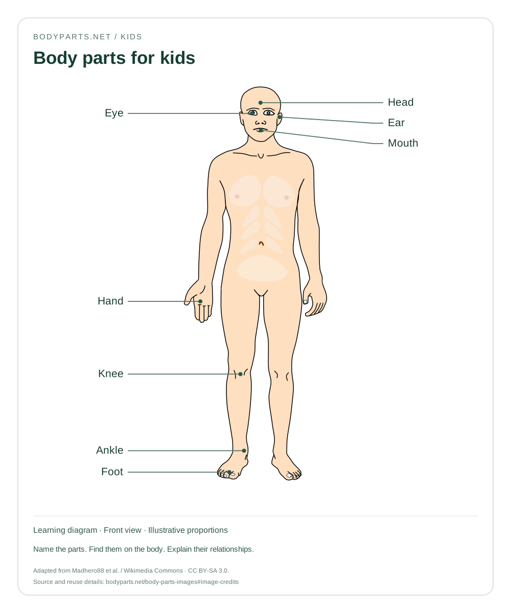
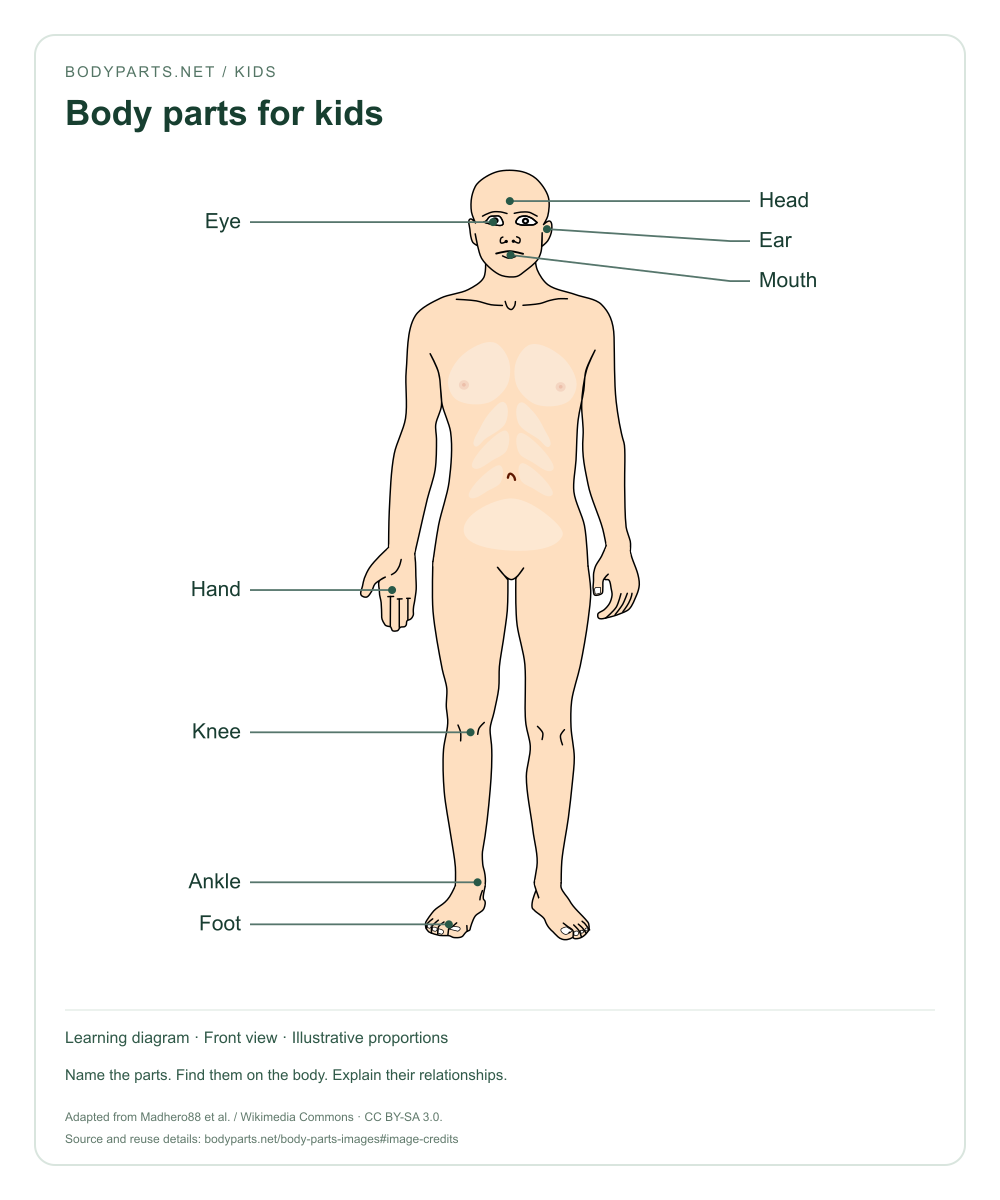
A first picture guide to eight familiar body parts. Point to each part, say its name and find the same part on your own body.
Read the anatomy guide →PICTURE LIBRARY / 18 EDUCATIONAL IMAGES
Download body parts pictures for kids, school lessons and anatomy education. Choose a labelled body parts diagram, a practice worksheet or a closer look at a region of the body.
PNG for everyday use
SVG for sharp prints
Open any image at full size, or download a PNG or SVG. Diagrams are 1000 × 1200 pixels and include their labels and credits. Keep the attribution when sharing or adapting them.
Kids · CC BY-SA 3.0
A first picture guide to eight familiar body parts. Point to each part, say its name and find the same part on your own body.
Read the anatomy guide →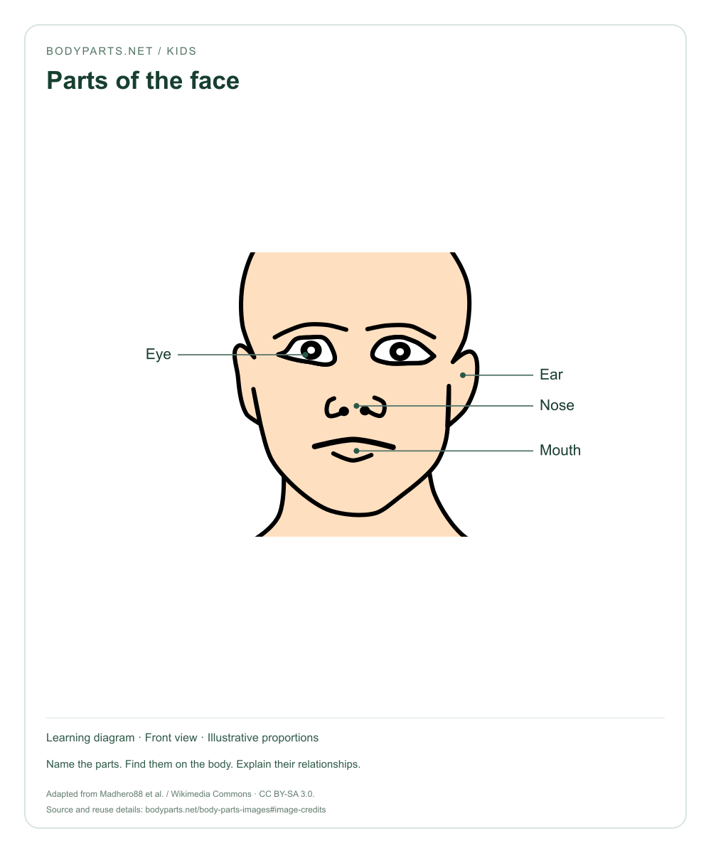
Kids · CC BY-SA 3.0
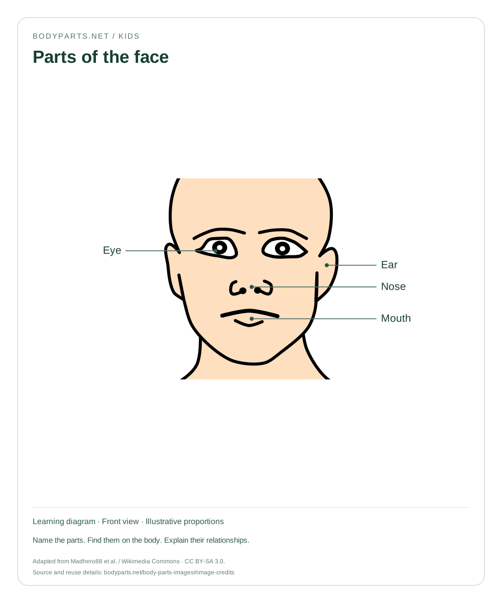
A close-up face diagram showing the eyes, ear, nose and mouth. Introduce facial vocabulary before exploring the smaller structures in each region.
Read the anatomy guide →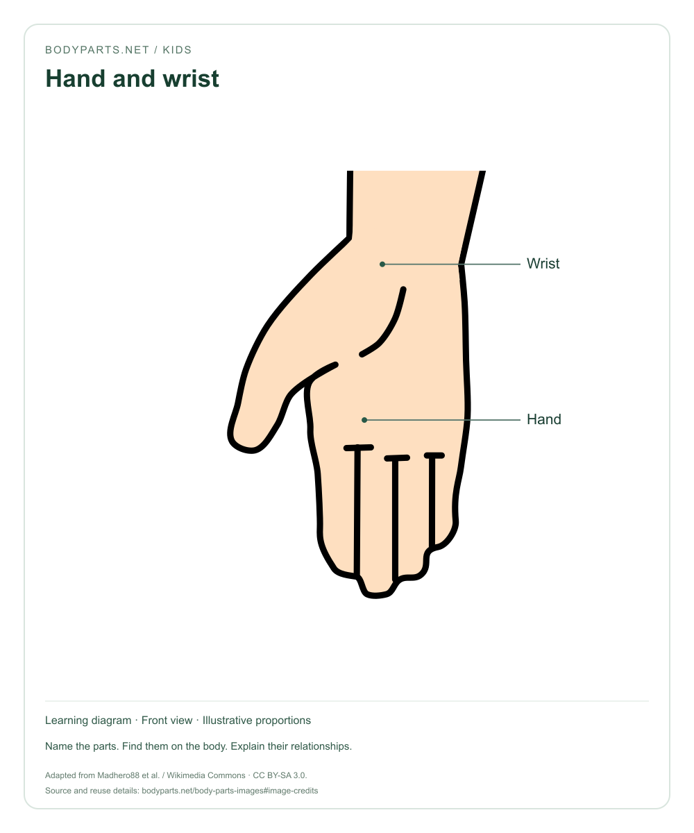
Kids · CC BY-SA 3.0
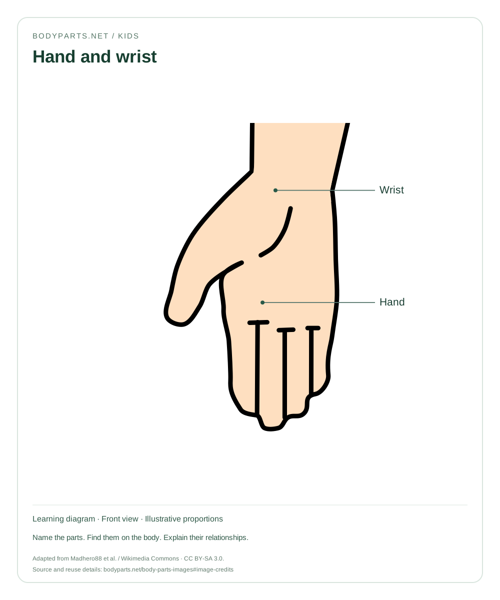
A close-up of the hand and wrist for early vocabulary practice. Use the accompanying hand guide to explore the palm, fingers, thumb and nails.
Read the anatomy guide →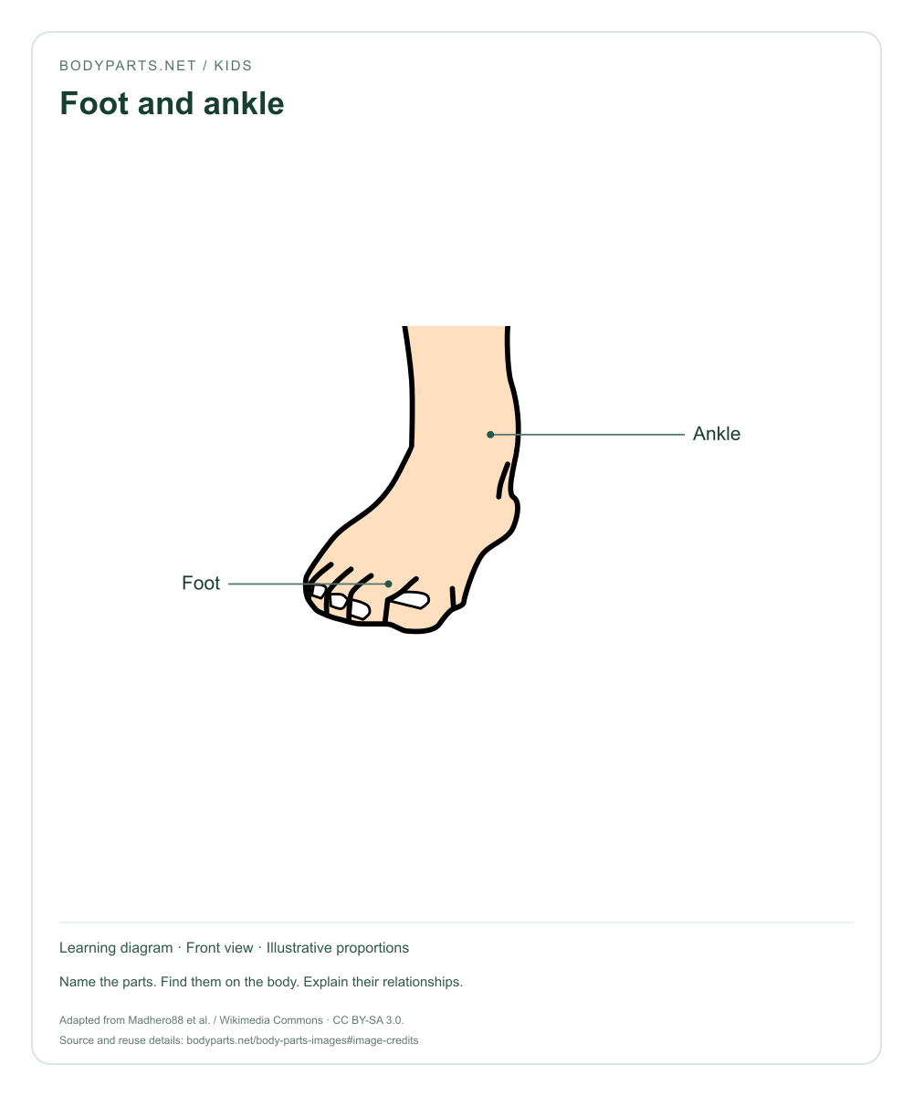
Kids · CC BY-SA 3.0
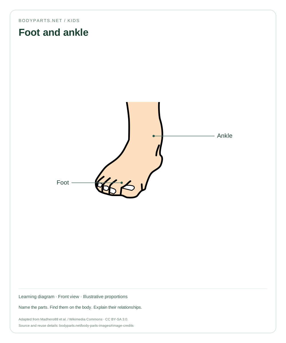
An enlarged foot-and-ankle picture for naming and comparison activities. The foot guide explains the heel, sole and toes in more detail.
Read the anatomy guide →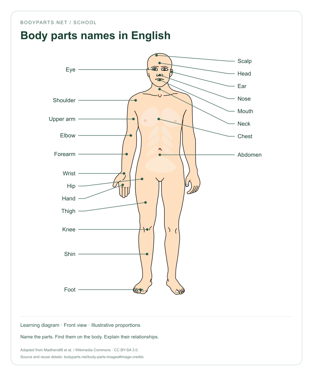
School · CC BY-SA 3.0
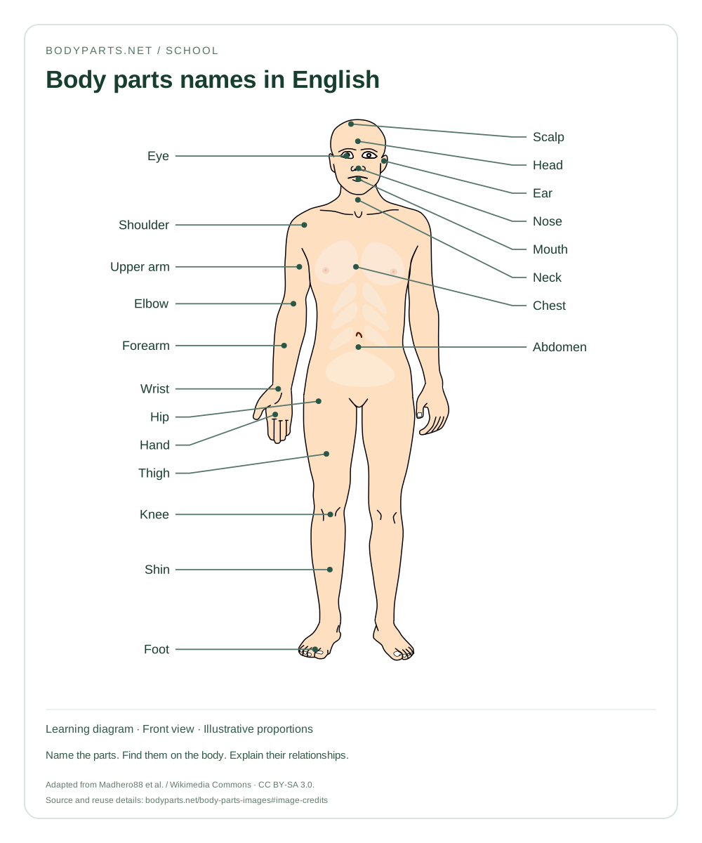
A vocabulary chart with 20 English body part names, arranged from head to toe. Use it for word recognition and classroom revision.
Read the anatomy guide →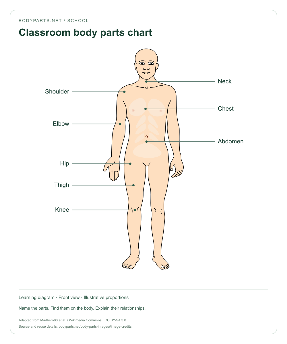
School · CC BY-SA 3.0
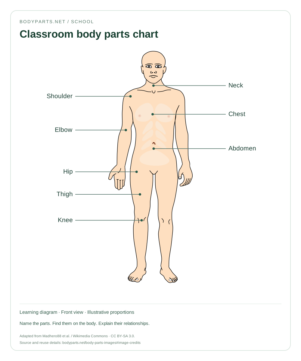
A regional anatomy chart for classroom discussion. Compare the torso, upper limbs and lower limbs, then describe where each named region is located.
Read the anatomy guide →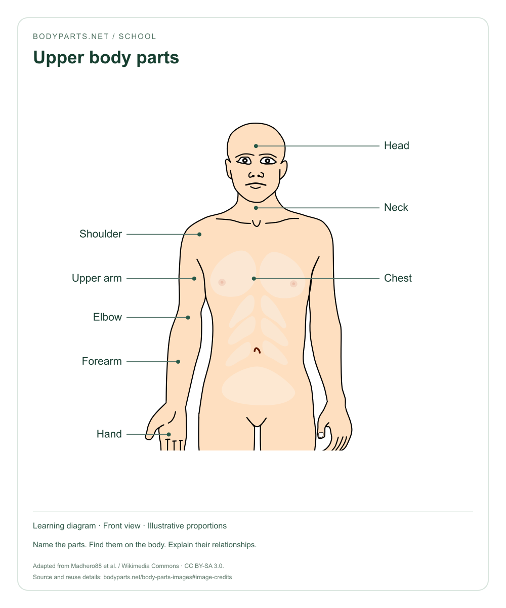
School · CC BY-SA 3.0
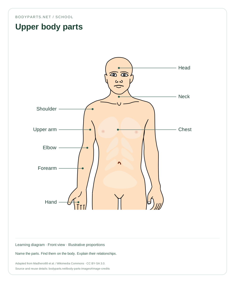
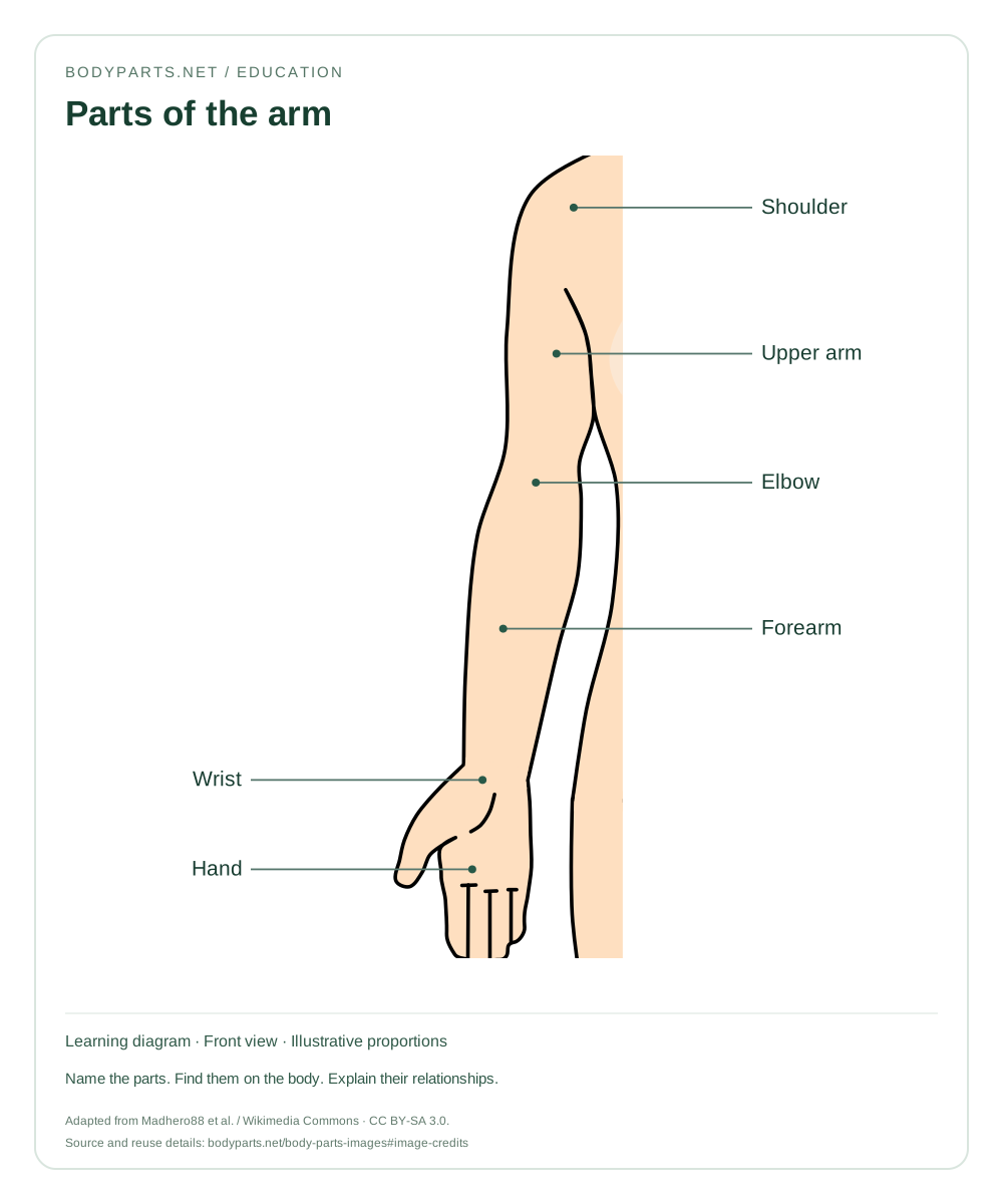
A closer view of upper-body regions. Follow the upper limb from the shoulder through the elbow and forearm to the hand.
Read the anatomy guide →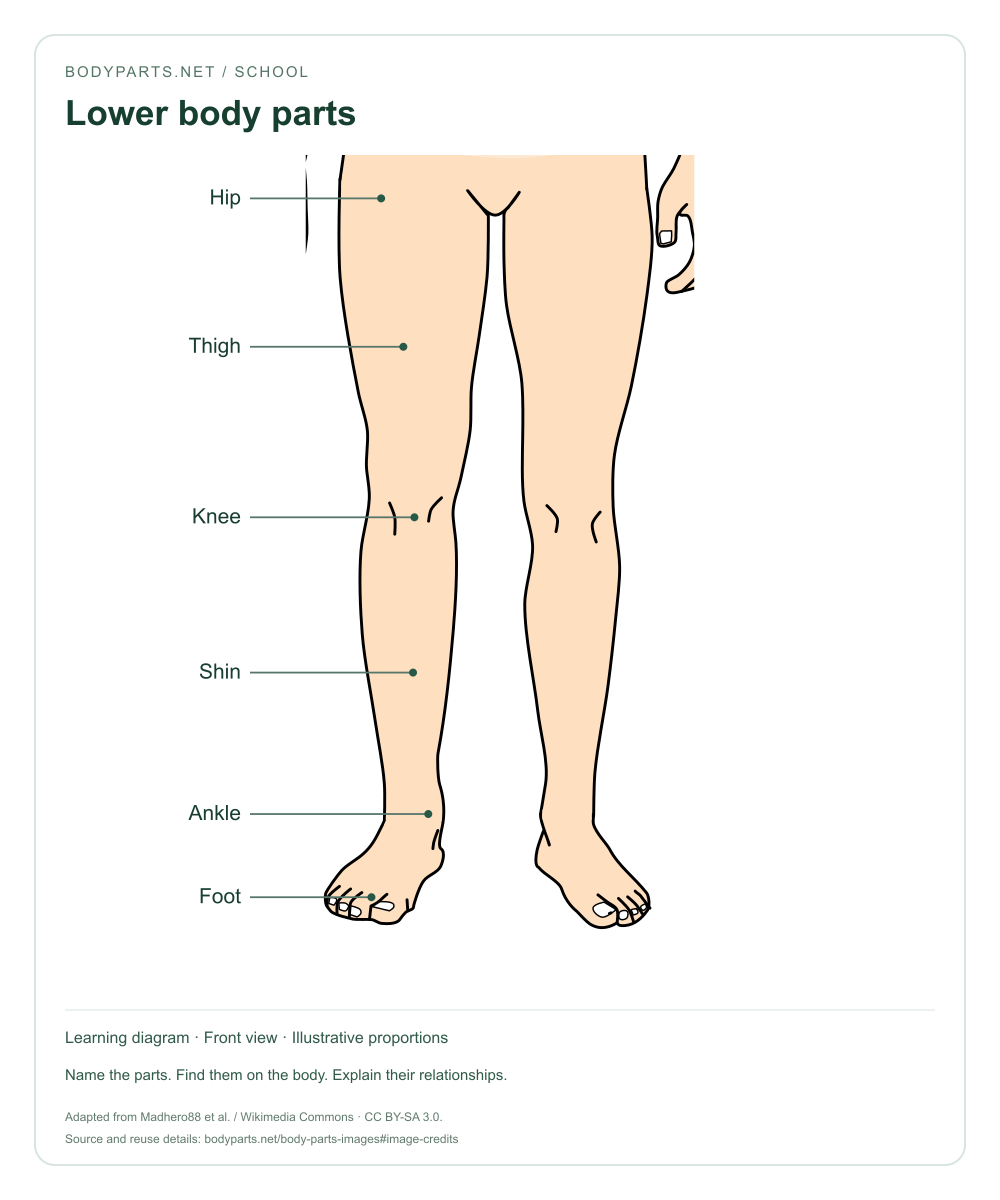
School · CC BY-SA 3.0
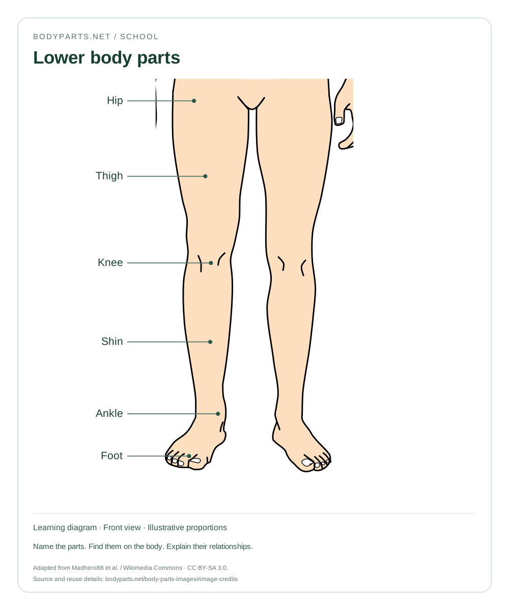
A lower-body chart for naming the hip, thigh, knee, shin, ankle and foot. Follow the labels in order from the pelvis toward the ground.
Read the anatomy guide →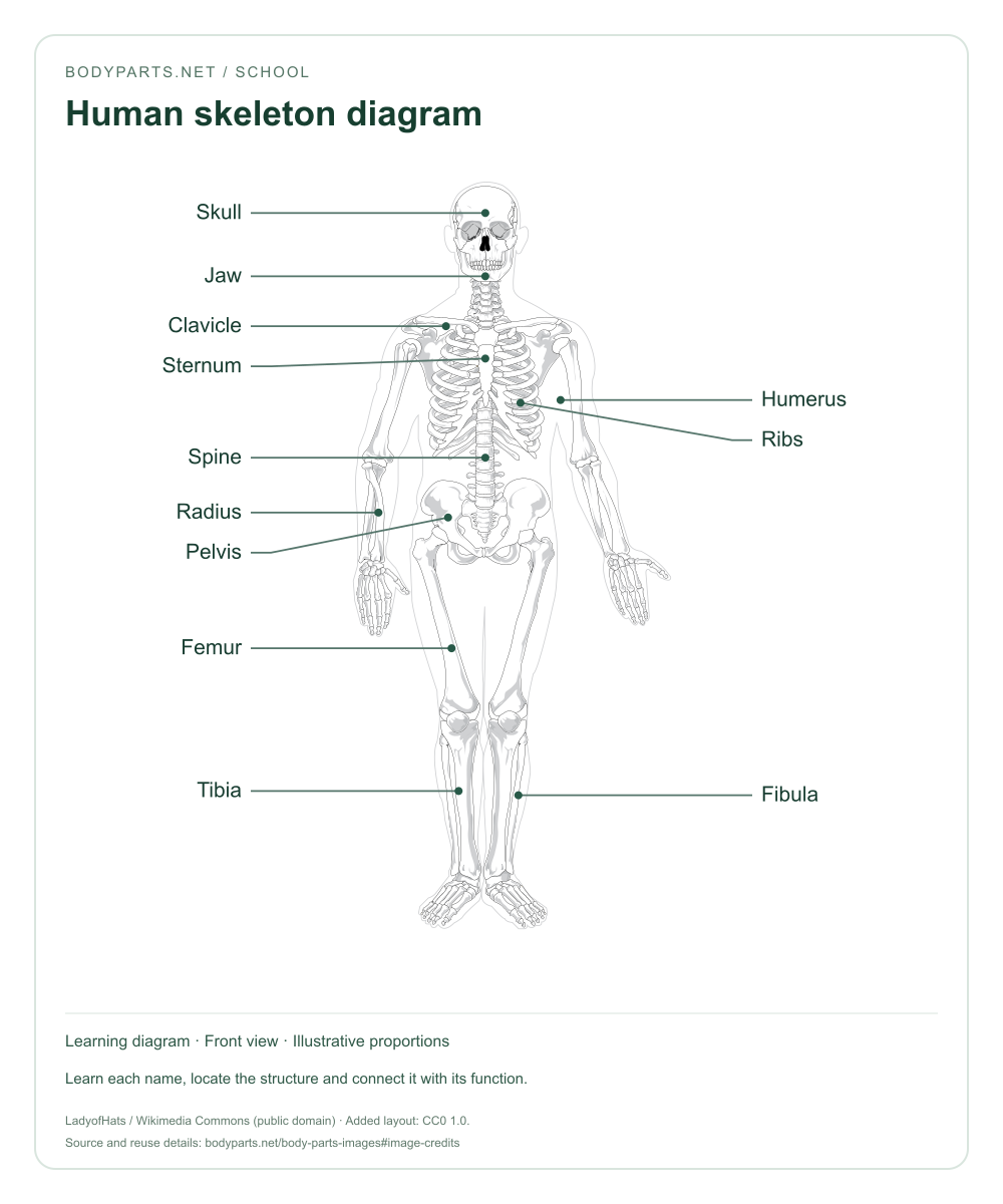
School · CC0 1.0
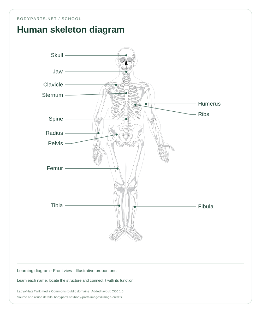
A labelled skeleton chart showing major bones and the vertebral column. Compare this internal framework with the external body parts chart.
Read the anatomy guide →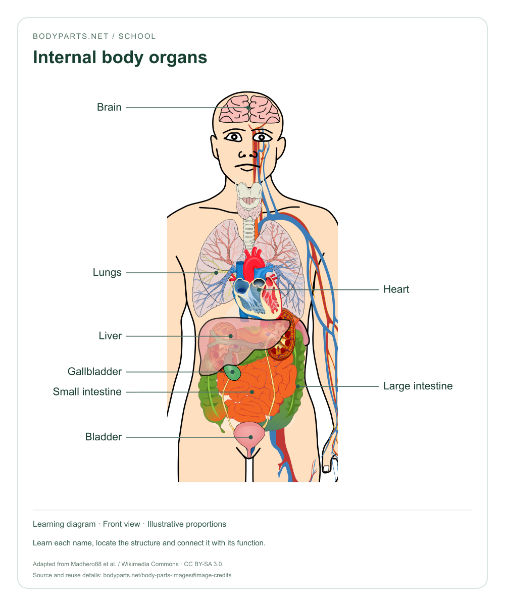
School · CC BY-SA 3.0
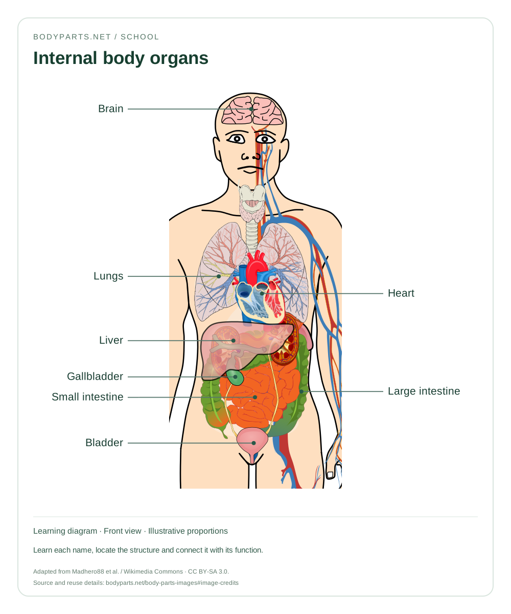
A cutaway overview of selected internal organs. This image introduces organ locations alongside, rather than instead of, external body part names.
Read the anatomy guide →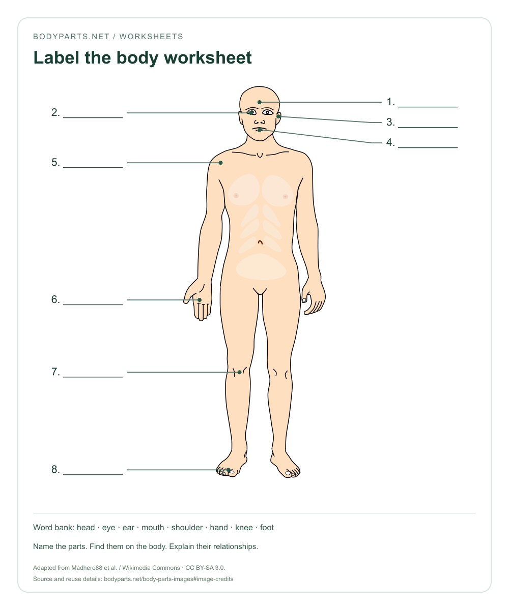
Worksheets · CC BY-SA 3.0
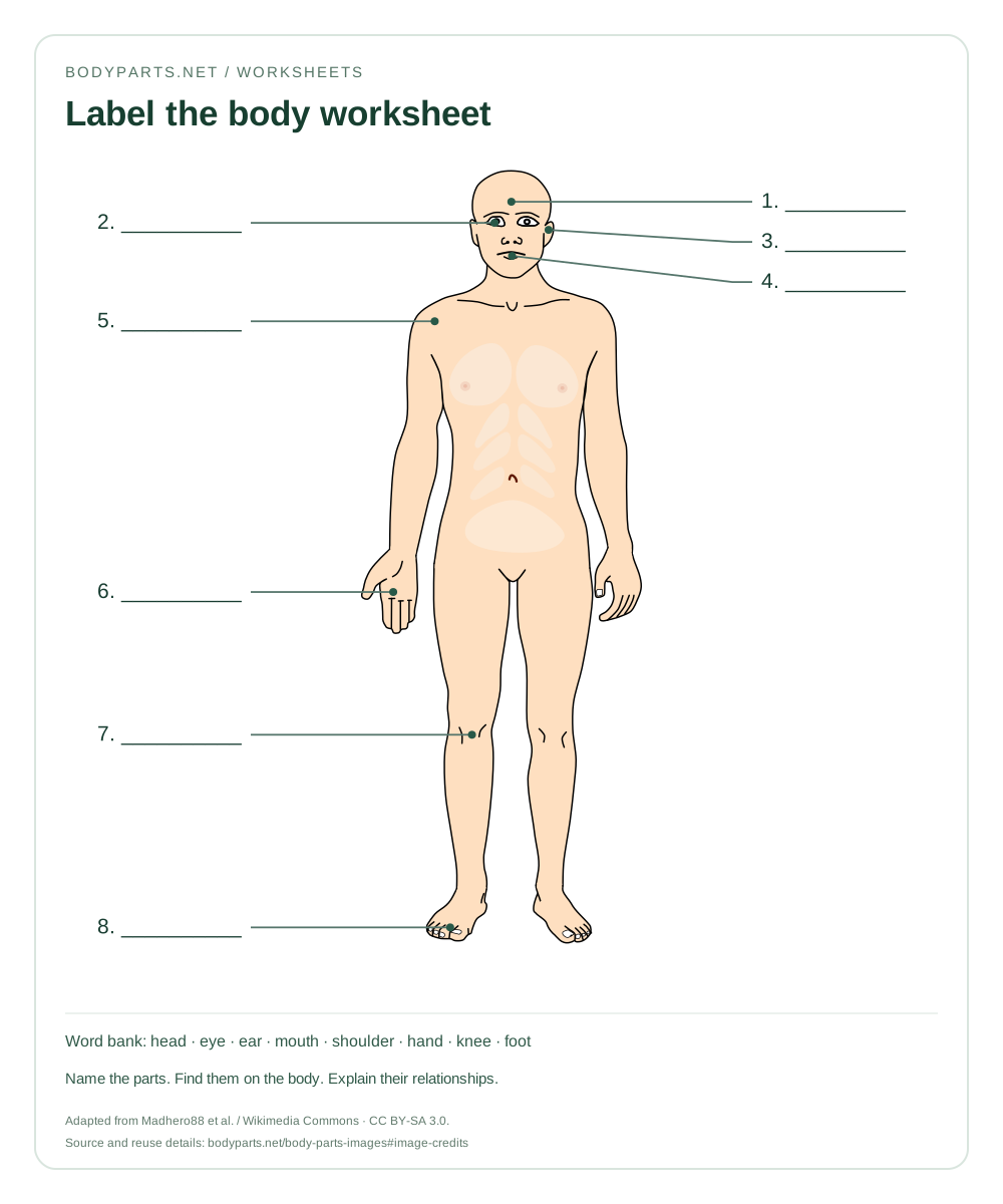
An unlabelled practice sheet with eight numbered answer spaces. Write the body part names, then check against the word bank beneath the diagram.
Read the anatomy guide →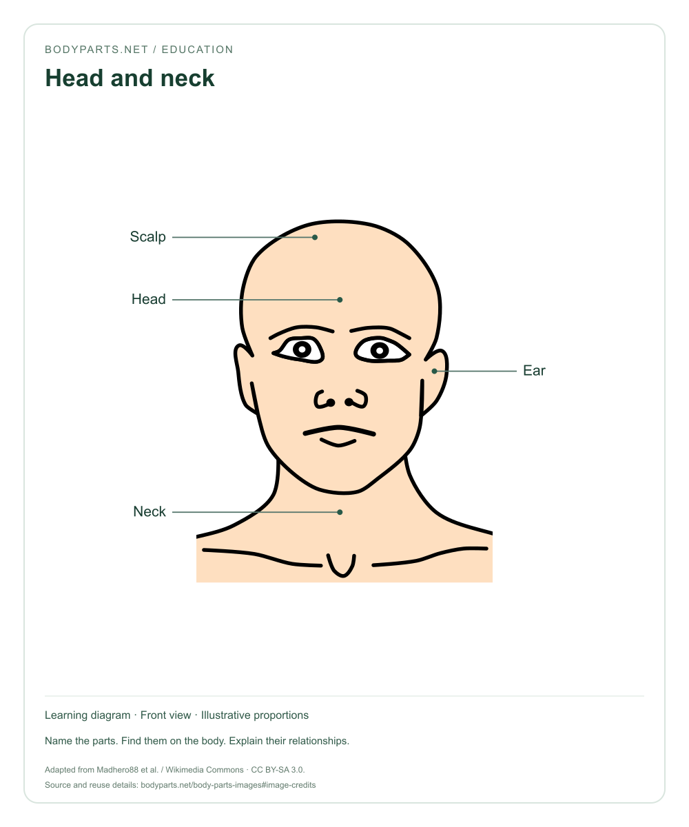
Education · CC BY-SA 3.0
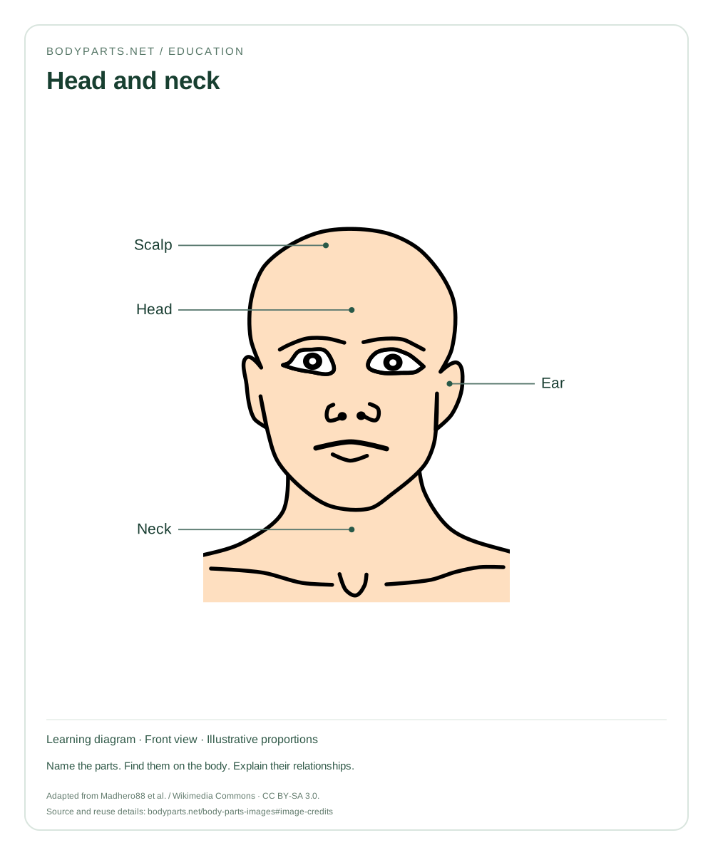
A head-and-neck reference image that distinguishes the scalp, head, ear and neck. Useful for introducing anatomical regions.
Read the anatomy guide →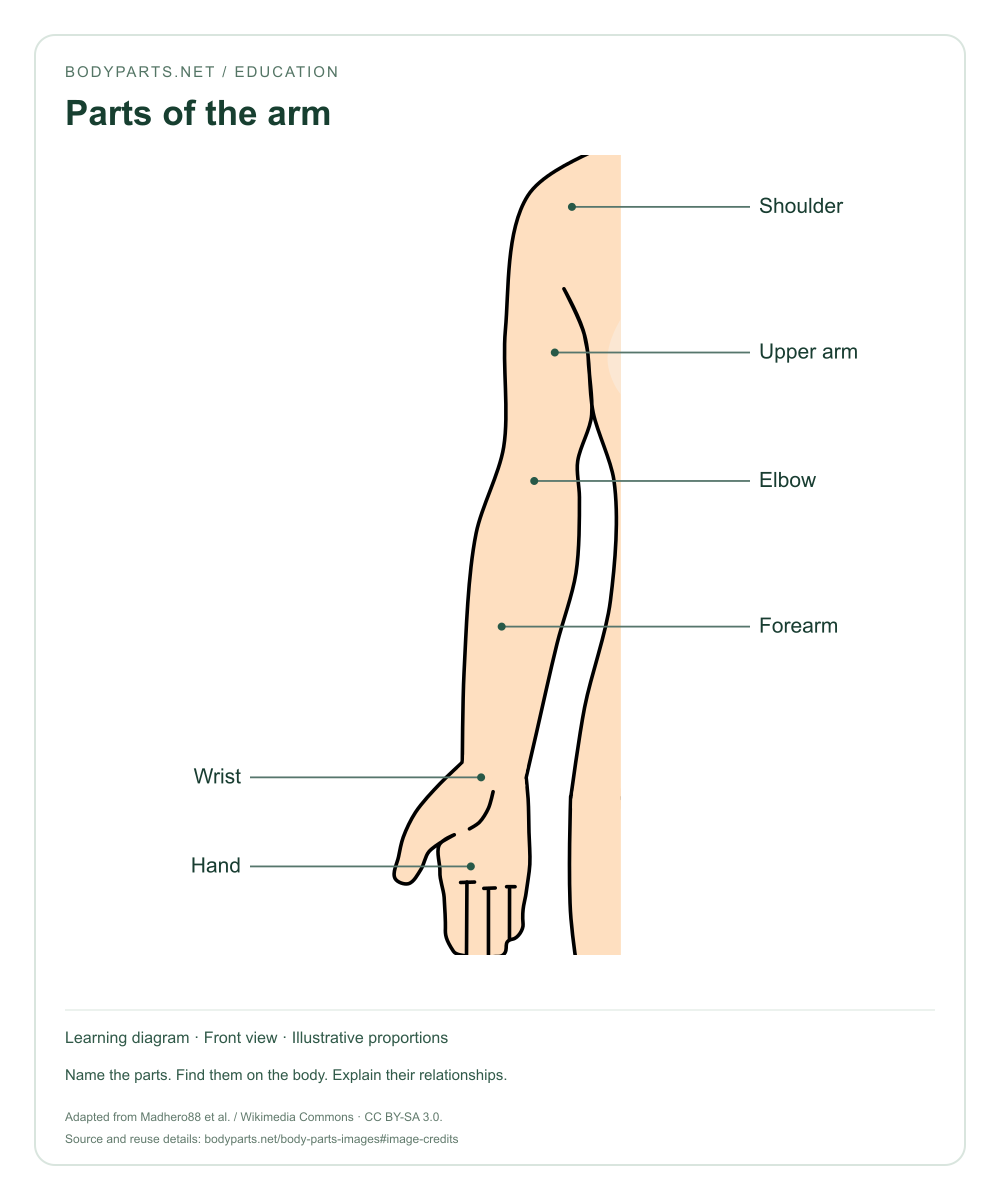
Education · CC BY-SA 3.0
A labelled arm image showing the sequence of regions from shoulder to hand. Compare the upper arm with the forearm.
Read the anatomy guide →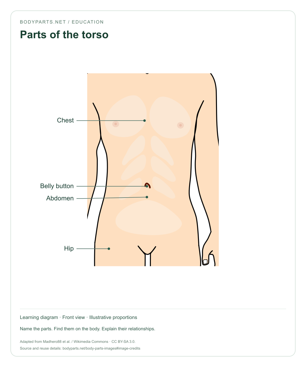
Education · CC BY-SA 3.0
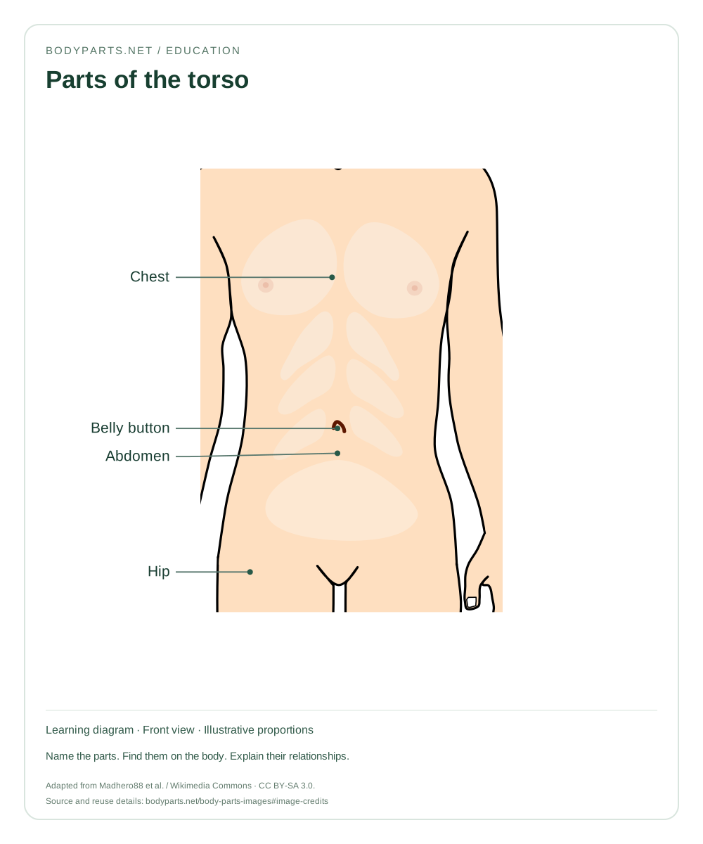
A torso diagram locating the chest, abdomen, belly button and hip region. Use it to distinguish external regions from the organs beneath them.
Read the anatomy guide →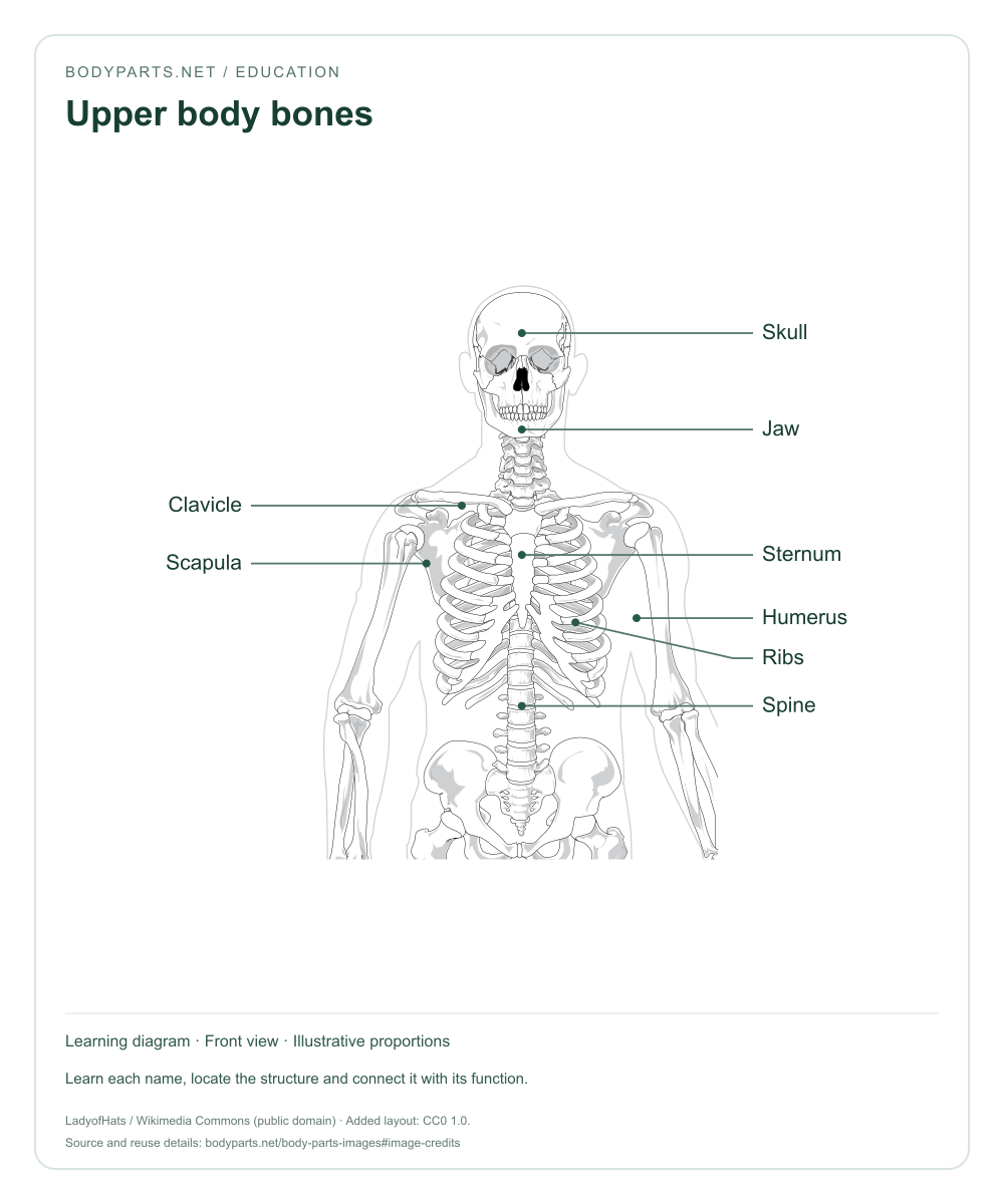
Education · CC0 1.0
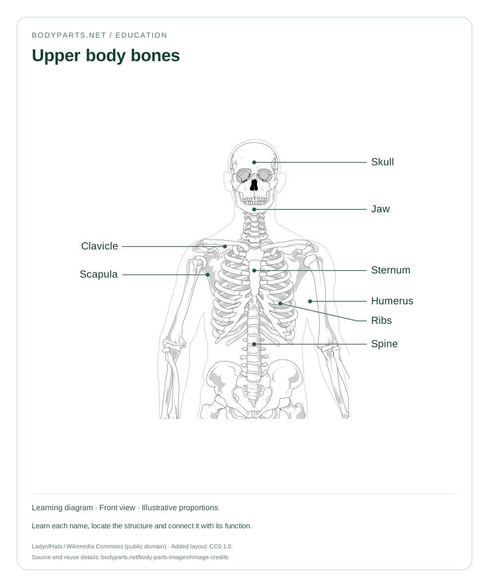
An upper-skeleton image for learning the skull, shoulder girdle, rib cage and upper-arm bone. Labels introduce the main anatomical names.
Read the anatomy guide →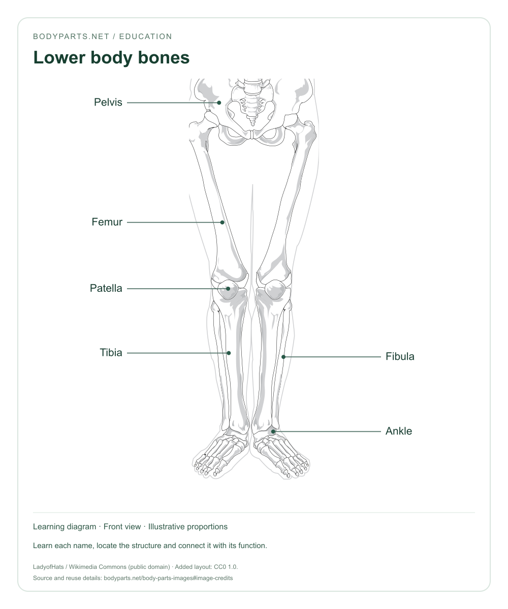
Education · CC0 1.0
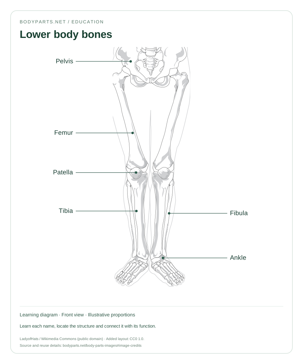
A bone diagram from pelvis to ankle. Compare the large femur with the tibia and fibula, and locate the patella at the knee.
Read the anatomy guide →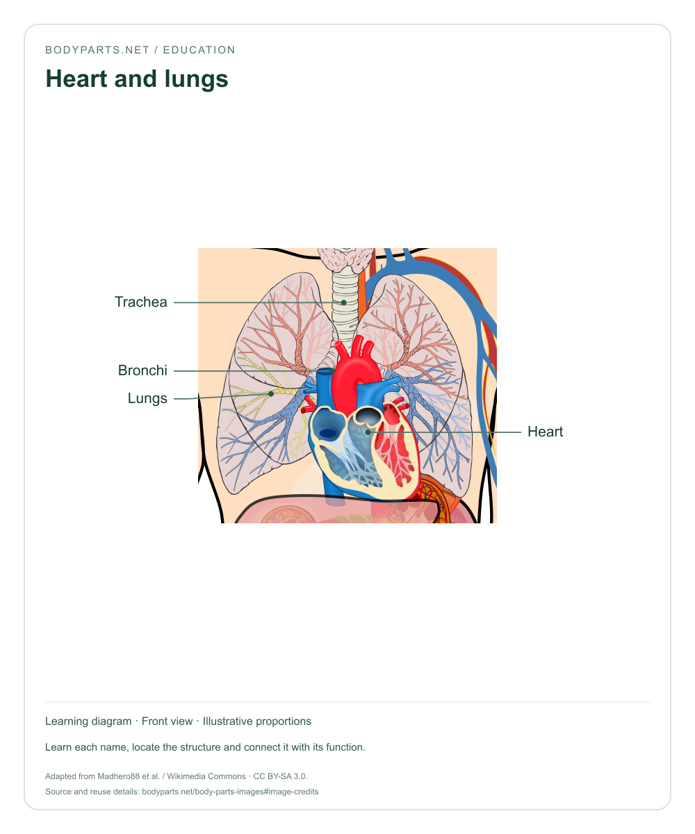
Education · CC BY-SA 3.0
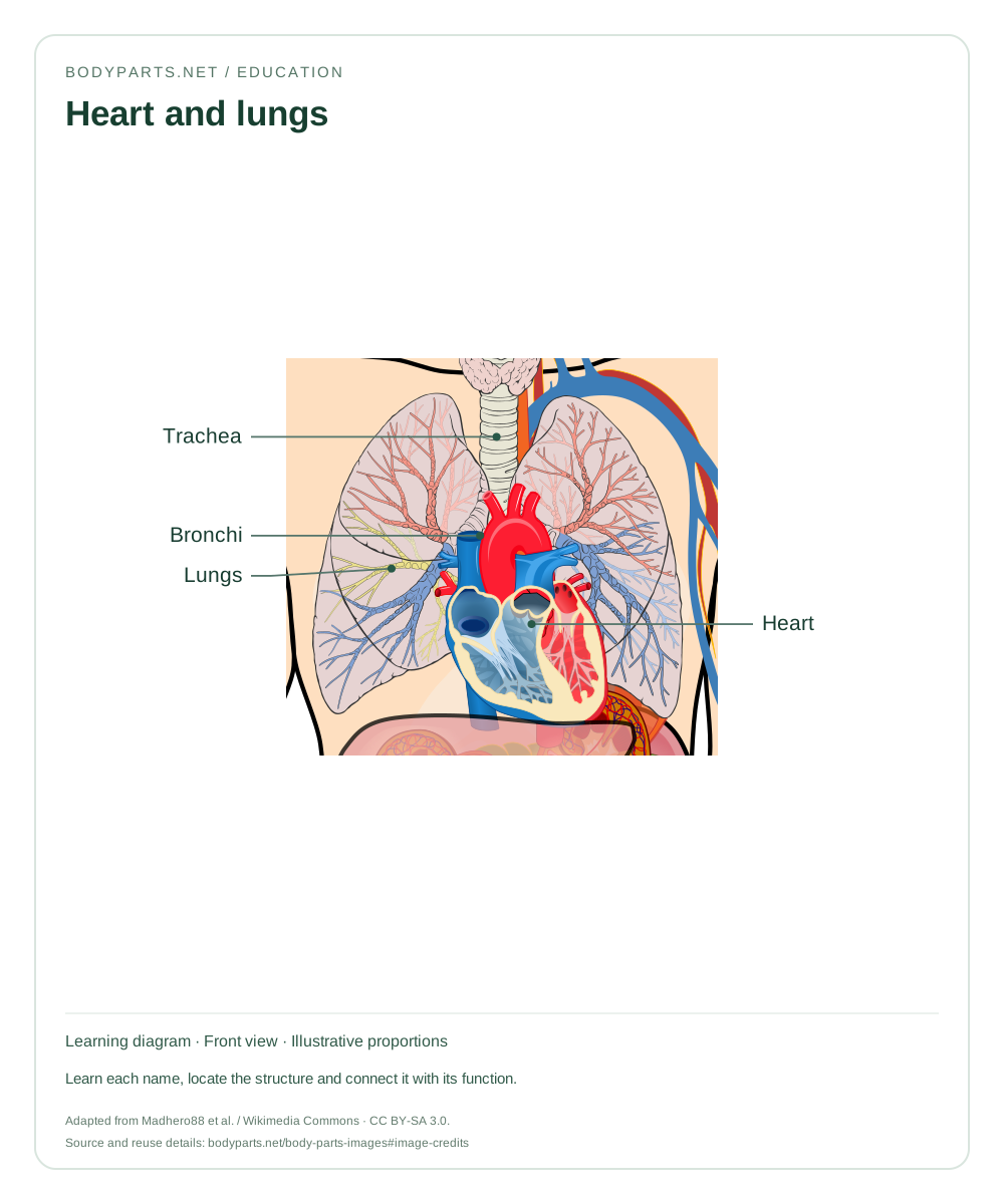
A chest-organ diagram showing the trachea, bronchi, lungs and heart. Discuss how breathing and circulation work together.
Read the anatomy guide →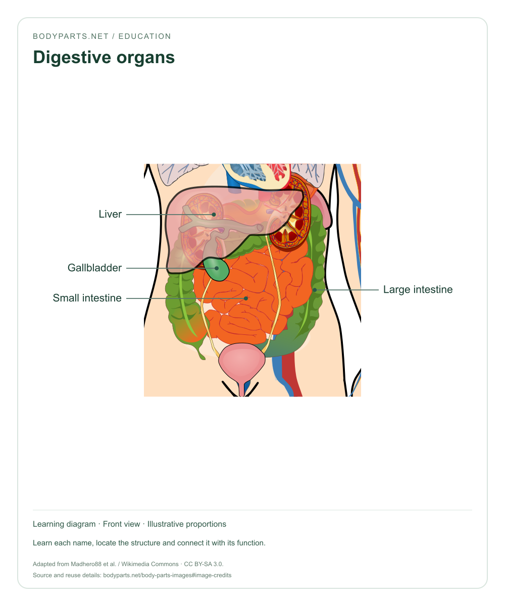
Education · CC BY-SA 3.0
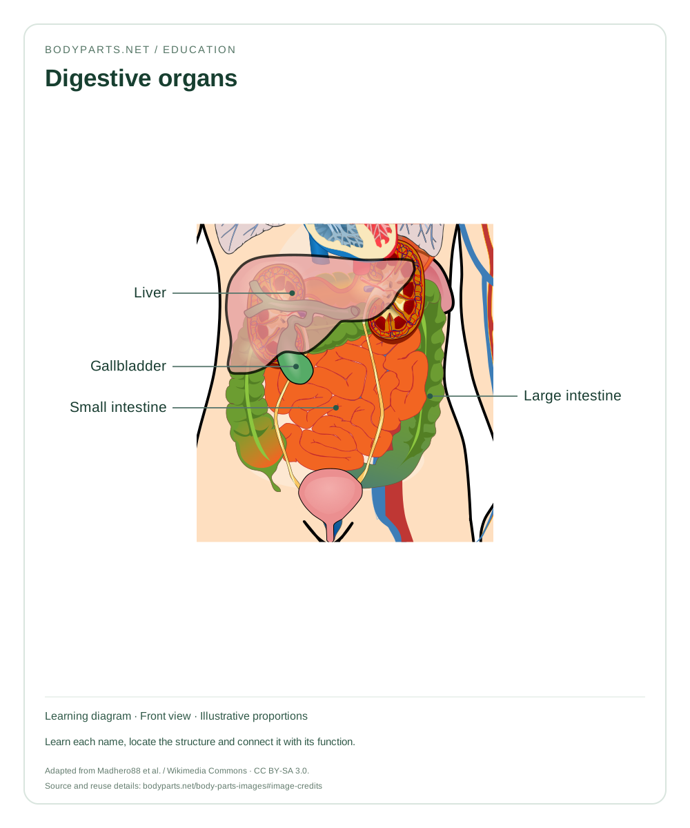
A labelled digestive-organ image showing the liver, gallbladder and intestines. Use it alongside the digestion study guides.
Read the anatomy guide →For younger learners, begin with a few familiar names and ask them to point to the matching region. For school lessons, pair a labelled image with the worksheet or compare an external diagram with the skeleton. For more advanced study, connect each named structure to its location, function and neighboring tissues in the anatomy guides.
These are simplified reference diagrams with adult proportions. They illustrate selected structures and do not represent every body type or a complete anatomical dissection. A front view shows the person's right on the reader's left.
Explore all body parts names →The external-body and organ illustrations are adapted from SVG Human With All Organs, assembled by Madhero88 with illustrations credited to LadyofHats (Mariana Ruiz Villarreal), ZooFari, Piom, Patrick J. Lynch and Mikael Häggström. These adapted diagrams are offered under Creative Commons Attribution-ShareAlike 3.0.
Skeleton illustrations use the public-domain artwork of LadyofHats (Mariana Ruiz Villarreal). Bodyparts.net added crops, labels, leader lines, worksheet numbering and page layouts. Our added layouts and labels for skeleton images are released under CC0 1.0. You may use the pictures for lessons and other purposes allowed by their licenses; retain attribution and apply the required share-alike terms to adaptations of CC BY-SA images. Full illustration attribution.